Presentation
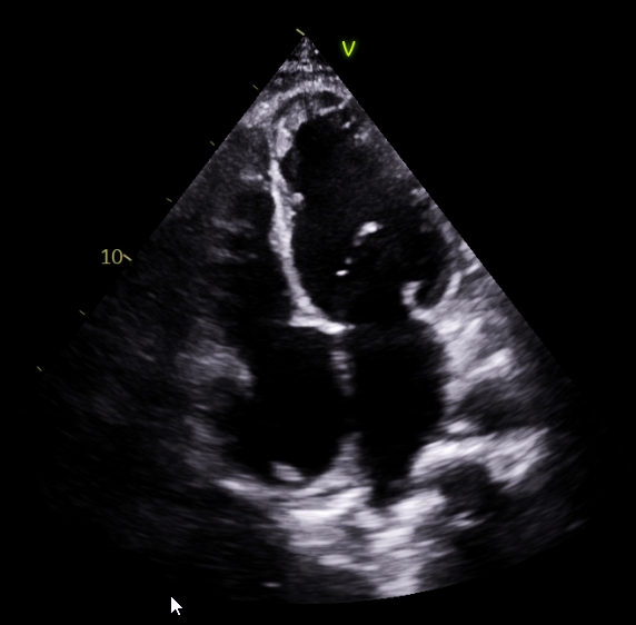
A man in his 30s was referred for pre-operative cardiac assessment before planned epigastric/umbilical hernia repair. Echocardiography unexpectedly demonstrated regional wall motion abnormality, mild left ventricular systolic dysfunction (LVEF 44%), Grade I diastolic dysfunction and an organized LV apical thrombus with minimal mobility.
He reported an episode of chest pain several years earlier that had been managed conservatively without further cardiac evaluation. This raised suspicion of a previously unrecognized anterior wall myocardial infarction with subsequent LV thrombus formation.
Investigations
Laboratory evaluation showed significant polycythemia, with haemoglobin of 19.9 g/dL and a packed cell volume of 54%. Leukocyte and platelet counts were normal. The reported workup included serum homocysteine and protein C testing; HRCT of the chest showed no significant abnormality.
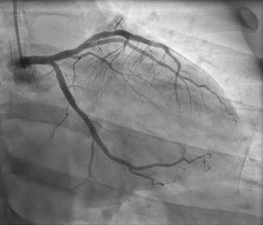
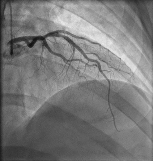
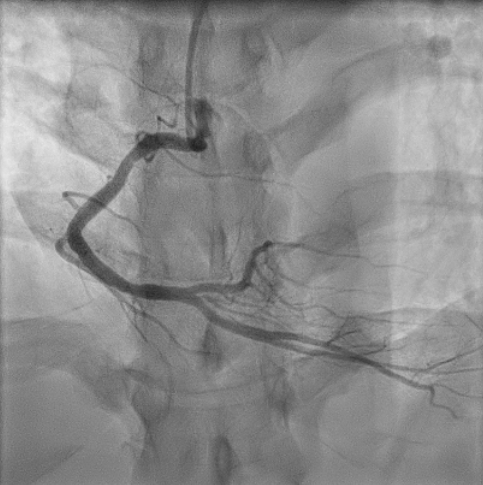
Coronary angiography subsequently showed minor coronary artery disease, with probable recanalized LAD disease described in the case report. The previous myocardial infarction remained a suspected explanation rather than a documented historical diagnosis.
Management and clinical course
The planned hernia operation was postponed because of the risk of systemic embolization. Cardiology, Hematology/Oncology and General Surgery coordinated further evaluation and treatment.
- Cardiac and haematological assessment. ECG, echocardiography, laboratory investigations and coronary angiography were undertaken, alongside evaluation of polycythemia and a possible pro-thrombotic state.
- Anticoagulation and monitoring. The patient received prolonged warfarin therapy with serial INR monitoring. Repeated sub-therapeutic INR measurements required several dose adjustments. Antiplatelet and statin therapy were also reported.
- Reassessment and surgical planning. Follow-up imaging documented an organized/stable LV thrombus. After completion of anticoagulation and clinical stabilization, warfarin was discontinued and elective hernia repair subsequently proceeded.
Clinical images
Outcome
No systemic embolic complications were reported during the documented clinical course. The LV thrombus was described as organized/stable on follow-up, and the patient remained clinically stable and asymptomatic. Elective hernia repair was eventually completed successfully.
The source report does not specify the total duration of anticoagulation or document complete thrombus resolution. The outcome is therefore presented as stabilization, rather than confirmed resolution.
Case reflections
- An incidental cardiac finding during pre-operative assessment substantially changed the surgical plan in this patient.
- Polycythemia added a potentially pro-thrombotic factor and prompted specialist evaluation.
- Repeated sub-therapeutic INR results made longitudinal monitoring and warfarin adjustment a central part of care.
- Coordination between cardiology, haematology/oncology and surgery supported eventual completion of the postponed operation.
Adapted from the author-supplied ADFHS 2026 case submission. Patient identifiers and exact dates have been omitted. Images are reproduced from the supplied report. This account describes an individual clinical course.
← Back to Clinical Cases